Imaging Anatomy: Chest, Abdomen, Pelvis - Imaging Anatomy
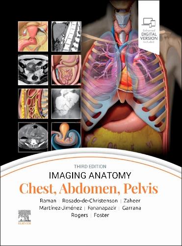
Imaging Anatomy: Chest, Abdomen, Pelvis - Imaging Anatomy
hardback
Published:
16 October, 2023

Popular Books by Siva P. Raman
View All Books by Siva P. Raman
Description
More Details
| Type | Book |
|---|---|
| ISBN13 | 9780443118005 |
| ISBN10 | 0443118000 |
| Number Of Pages | 1192 |
| Item Weight | 3720 g |
| Publisher / Reseller | Elsevier Health Sciences |
| Format | hardback |
| Edition | 3rd edition |
Author's Bio
Dr. Siva Raman is a board-certified radiologist who gained his subspecialty expertise in thoracoabdominal imaging during a fellowship at Stanford University, and intern residency at UC Davis Medical Center. He attended Johns Hopkins University Medical School. Dr. Raman is a radiologist at Bay Imaging Consultants in Walnut Creek, California, with a subspecialty focus on thoracoabdominal imaging Melissa L. Rosado-de-Christenson, MD, FACR, FAAWR, is Attending Radiologist at the Division of Cardiothoracic Imaging in the Department of Medical Imaging for Banner - University Medical Group Tucson,. She is also Professor of Medical Imaging at University of Arizona College of Medicine - Tucson, in Tucson, Arizona Dr. Atif Zaheer has a specialty focus on CT, MR, and US of the abdomen and pelvis. He is Professor of Radiology, Oncology and Medicine at The Johns Hopkins University School of Medicine in Baltimore, Maryland. Dr. Santiago Martínez-Jiménez is with the Department of Radiology at Saint Luke's Hospital of Kansas City and is Professor of Radiology at the University of Missouri-Kansas City School of Medicine, in Kansas City, Missouri. He's a board-certified practicing radiologist who specializes in cardiothoracic radiology Dr. Ghaneh Fananapazir is a radiologist with specialization in abdominal radiology. He is Professor of Radiology at the University of California - Davis, in Sacramento, California. Fananapazir's clinical focus includes nonvascular interventional procedures, advanced US, CT and MR techniques, vascular imaging, and abdominal organ transplantation (kidney, liver, pancreas). In addition to his clinical duties, Dr. Fananapazir is passionate about medical education and research, particularly relating to kidney transplantation, and has authored textbooks and peer-reviewed research in highly impactful medical journals. Sherief H. Garrana, MD, is a clinical assistant professor of radiology with the Division of Thoracic Imaging in the Department of Radiology at NYU Langone Health in New York, New York. Dr. Douglas Rogers is an Associate Professor in the Department of Radiology and Imaging Sciences at the University of Utah School of Medicine in Salt Lake City, Utah. Rogers is a board-certified Diagnostic Radiologist who specializes in abdominal imaging with a focus in gynecology. Dr. Bryan Foster is a body imaging radiologist with a subspecialty focus on oncologic, hepatobiliary, pancreatic, prostate, and small bowel imaging. Dr. Foster is the Professor of Radiology at Oregon Health & Science University in Portland, Oregon, and performs complex image-guided biopsies; he also performs CEUS imaging and US elastography. Dr. Foster is skilled in all facets of imaging including radiography, fluoroscopy, ultrasound, CT, and MRI. His interests include oncologic, hepatobiliary, pancreatic, prostate, and small bowel imaging. Dr. Foster has recently started performing contrast-enhanced ultrasound imaging (as the only site in Oregon) and US elastography. He is one of two radiologists that perform MRI-guided prostate biopsies (also only site in Oregon). Recently he was selected as an Honored Educator for the Radiological Society of North America.